Imaging the activity of large populations of neurons within the brain could be critical towards understanding the function of neuronal circuits. Spatial Light Modulator (SLM) microscopy is a simple holographic method that allows one to simultaneously image activity of neurons across large spatial areas or in multiple layers (L2/3 and L5) of the mouse cortex in vivo. This approach allows simple and rapid multiplexing, is low cost and could be used as a general method neuronal activity in three dimensions across multiple areas of the brain.
TOP (A) Optics diagram for the setup of SLM. (B) Illustration of axial dual plane imaging, where two planes at different depths can be simultaneously imaged. (C) Illustration of lateral dual plane imaging, where two adjacent fields at the same depth can be simultaneously imaged.
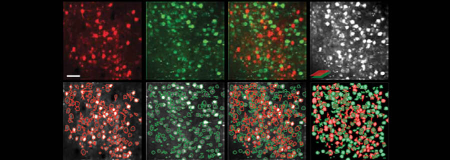
BOTTOM: Axial Dual Plane in-vivo Functional Imaging of Mouse V1 at Layer 2/3 and 5 (A) / (B) Top panel, temporal standard deviation images of the sequential single plane recording (10 fps) of mouse V1 at depth of 170 μm (layer 2/3) and depth of 500 μm (layer 5) from the pial surface. The images are falsecolored. Bottom panel, spatial component contours overlaid on the top panel. Scale bar, 50 μm. (C) Arithmetic sum of (A) and (B). (D) Top panel, temporal standard deviation image of the simultaneous dual plane (10 fps) recording of the two planes shown in (A) and (B). Bottom panel, overlaid spatial component contours from the two planes.